|
|
 |
|
||||
|
Teaching File Series |
Main |
|
||||
|
M R I - Division |
||||||
| Click on the images to view them closely | ||||||
|
||||||
|
||||||
|
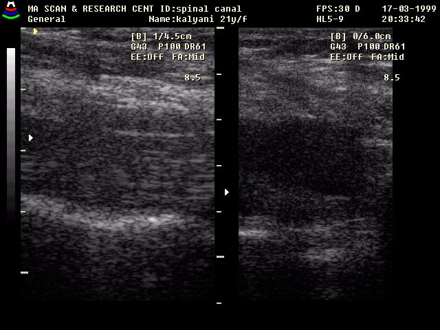
Complaint
: 21 y female with severe lower backache |
||||||
|
|
|
|
||||
|
Teaching File Series |
Main |
|
||||
|
M R I - Division |
||||||
| Click on the images to view them closely | ||||||
|
||||||
|
||||||
|
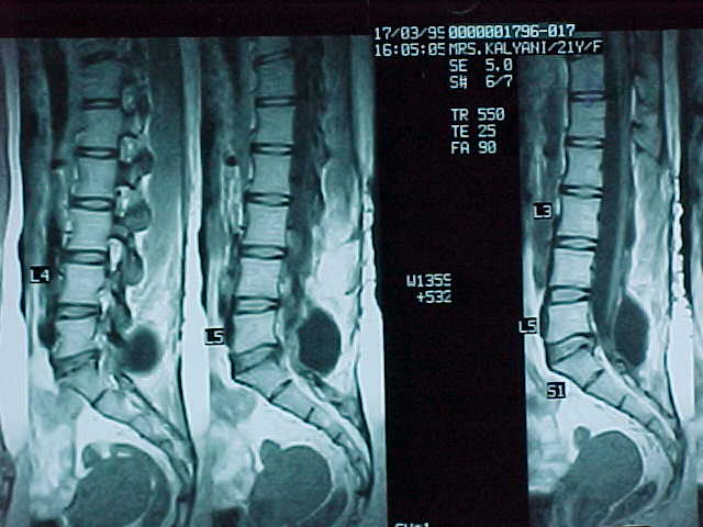
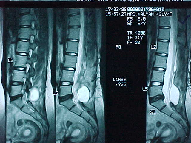
Complaint
: 21 y female with severe lower backache |
||||||
![]()
©2000 Radiologyworld.com