|
|
|
| |||||||
|
Teaching File Series |
Last
|
| |||||||
|
M R I - Division | |||||||||
| Click on the images to view them closely | |||||||||
| |||||||||
| |||||||||
|
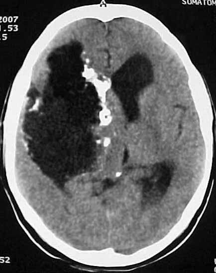
Complaint : 40Y, Female c/o seizures on and off since 14 years.
CT: Large lobulated cystic mass with peripheral calcification in the right temporo-frontal region compressing right lateral ventricle & obstructive dilatation of left lateral ventricles.
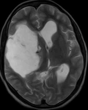
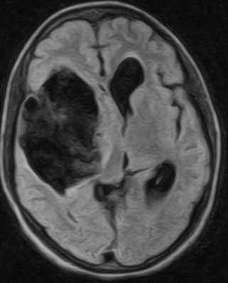
MRI :
Appears hypointense on T1W,
hyperintense on T2W & DWI. On ADC hypointense with incomplete suppression
on FLAIR. DISCUSSION : Congenital Epidermoid cyst arise from ectodermal inclusion during neural tube closer. Acquired Epidermoid cyst is uncommon and as a result of trauma. Epidermoid cyst is most common congenital intracranial tumor. 0.2 to 1.8 % all primary intracranial tumor. It grows by progresive desquamation with conversion to keratin / cholesterol crystals. It is soft pliable, conforms to shape of adjacent local structures / spaces. Presents at 20 to 60 year with peak at 40 years. Most commong symptom headache, Cranial nerve 5, 7, 8 neuropathy. Seizures if in sylvian fissure / temporal lobe. May remain clinically silent for many years. 90% intradural, primarily in basal cisterns. CP angle 40 to 50%. Parasellar / middle cranial fossa 10 to 15 %. On CT scan apears isodense to CSF in >95% & calcification in 10 to 25%. On MRI slightly hyperintense to CSF on T1 W & isointense to CSF on T2 W. Usually on FLAIR doesnot completely null. DW shows restricted diffusion and yields high signal & ADC isointense to brain parenchyma. CSF like mass imaging and shows restricted diffusion is diagnositc of epidermoid. REFERENCE : Osborn AG, Blaser SI, Diagnostic Imaging, Brain 1st edition, 2004. SUBMITTED BY: DR. M.ADINARAYANA RAO. MD. DR. SUNIL ALMALE. DMRD.
| |||||||||