CLINICAL FEATURES : 60y female
presented with pain in the knee joint.
SONOGRAPHIC
FINDINGS : Longitudinal sectional study (H.R.S)of medial
condyles of tibia & femur including joint space shows (Image 1)
small bony out growths (osteophytes) at junction of articular surface
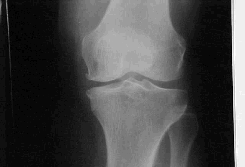
and condyles along with narrowing of joint space.Compare these
sonographic osteophytes with X-ray (Image 2). See the normal smooth
outlines of condyles ,articular surface with out osteophyte in normal
knee for comparison( image 3).
DIAGNOSIS :
Sonographic diagnosis of O.A.KNEE.
DISCUSSION
: High resolution (H.R.)sonographic diagnosis of oasteoarthritis of
knee is easy, once familiar with normal sonographic morphology
of knee joint. Narrow joint space, presence of osteophytes are
hallmark for osteoarthritis .H.R.sonography is not only useful in diagnosis but
also reveal bursitis/synovial effusions ,if any.
SUBMITTED
BY : Dr.M.Adinarayana raoM.D.(Radio-Diagnosis)