Clinical details : 20
female with G2L1
7TH MONTH
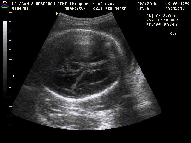
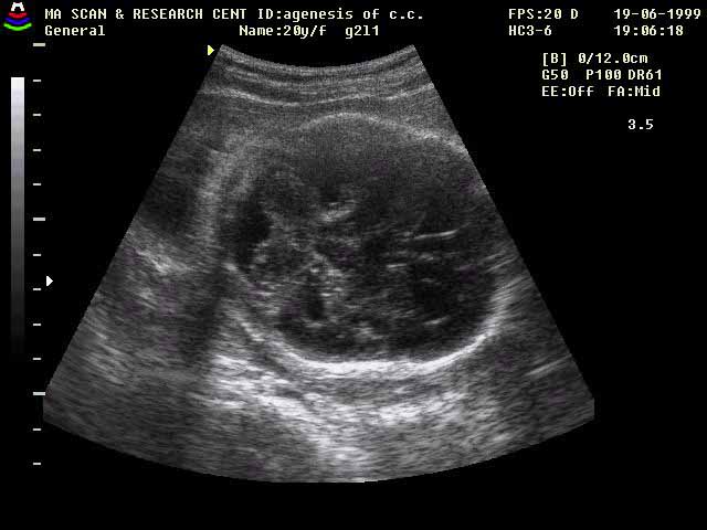
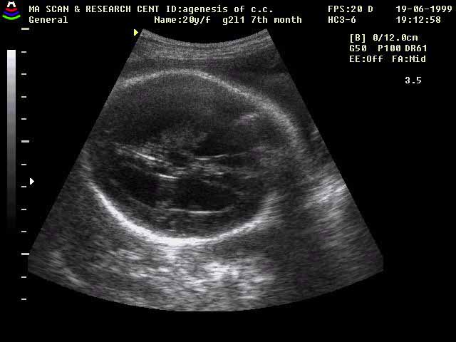
Findings :
3rd & lateral ventricles are dilated.
Medial wall of lateral ventricles are separated from mid line.
3rd ventricle is at high level.
Cisterna magna is normal.
Inter
hemispheric fissure is touching the 3rd ventricle.
Diagnosis :Agenesis of Corpus
Callosum.
Discussion :
A defective lamina reunions
results in either partial or complete
agenesis of corpus callosum. Complete agenesis is most
common. Absence of Corpus Callosum shows widely separated lateral ventricles with parallel medial
walls & enlarged, elevated 3rd ventricle. Abnormal 'radiating gyral ' pattern in the
Mesial hemispheres, best
seen in the latter part of 3rd trimester.
Common associations include inter hemispheric cyst, Intracranial
Lipoma, encephalocele,
Dandy-walker malformation, chary II malformation, Aquductal
Stenosis, Trisomy 13 &18. Agenesis of Corpus Callosum in the absence of other anomalies may be asymptomatic.
Aicardi syndrome is characterized by female with agenesis of Corpus
Callosum ocular
abnormalities & infantile spasms.
Reference :
RCNA vol:28 .,
Diagnostic Neuro Radiology by ANNE G. OSBORN.
Submitted by :
Dr.M.Adinarayana Rao. M.D. & Dr. C. Savithri. M. D.(GYN&OBG.)