|
Complaint : 36Y,
Male bilateral lower limb weakness, tingling & parasthesia from 6 months. No H/O Trauma.
Investigation:
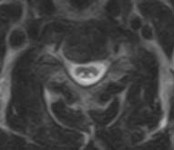
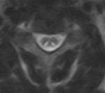
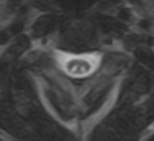
MRI Mid cervical spine, T2 Wt. Images of Axial sections.
FINDINGS:
MRI : Abnormal T2 hyper intensity confined
to dorsal columns with an inverted 'V' configuration or inverted rabbit
ears
DIAGNOSIS:
Vitamin B12 deficiency / sub acute combined degeneration of the
cord.
DISCUSSION
T2 hyper intense confined to
dorsal columns highly suggestive of Vitamin B12 deficiency.
Confirmation with laboratory
data decreased plasma B12 levels.
Differential Diagnosis :
Spinal cord infarction
-
Hyperacute
presentation, motor > sensory symptoms
-
Predominantly
ventral cord or central gray matter signal changes
Spinal cord contusion
-
Cord swelling,
T2 hyperintensity +/- hemorrhage
-
Associated
fracture, soft tissue injury
-
History,
clinical findings enable diagnosis
Inflammatory demyelination
-
Multiple
sclerosis or acute disseminated encephalomyelitis (ADEM)
-
Lesions more
focal, patchy than B12 deficiency, do not show specificity for
lateral or dorsal columns
-
Characteristic
clinical presentation
Infectious myelitis
-
HIV vacuolar
myelopathy, Varicella-Zoster/Herpes, Lyme disease
-
Imaging
findings may be identical to B12 deficiency
-
Clinical,
laboratory findings help distinguish
Acute transverse myelitis
-
Acute
(non-traumatic) presentation => diffuse multisegmental cord
hyperintensity, swelling
-
Idiopathic or
known etiology; clinical and laboratory findings may help
distinguish cause
-
Increased CSF
protein, pleocytosis, +/- oligoclonal bands (28%)
Special features to
remember
-
Spinal cord
symptoms appear first with motor (spastic paraparesis, gait
unsteadiness) .
-
and sensory (paraesthesias,
absent reflexes, loss of joint position sense and vibration sense).
-
Treatment
arrests degenerative process but does not restore destroyed neural
fibers.
-
Imaging changes
may not completely resolve.
-
Avoid N2O
anesthesia in vulnerable patients.
-
Common in
strict Vegetarians
REFERENCE : Ross, Brant-Zawadzki.Moore, Diagnostic Imaging,
Spine 1st edition, 2004.
SUBMITTED BY:
Academic division, 'MA' Advanced Diagnostic & Research Centre, Guntur, AP. INDIA.
DR. M.ADINARAYANA RAO. MD.
DR. G.GOWRI SEKHAR . DNB.,DMRD.
DR. D. PRASAD REDDY. DMRD.
|