INTRODUCTION
SONOGRAPHIC TECHNIQUE
NORMAL SONOGRAPHY OF MAXILLARY
SINUSES.
ABNORMAL SONOGRAPHY OF MAXILLARY
SINUSES
EXAMPLES
CONCLUSIONS
INTRODUCTION :
Maxillary sinuses are periodically filling
with air and conistantly exposing to virus bacteria
and allergic
chemicals. Hence prone to sinusitis ( Fluid-air layer)/ mucosal thicking.
To detect maxillary sinuses pathalogy x-ray is less sensitive & less
specefic. Radiation +.
CT scan is more sensitive & more specific. Radiation +. Price is more
Ultrasound is more sensitive & more specific than X ray. But less price
than CT scan. No radiation
SONOGRAPHIC TECHNIQUE:
EQUIPMENT :Higher end equipment with Lenear 7.5Mhz ,10Mhz probes &
color doppler.
Examine the patient maxillary sinuses on both siting & lying positions.
Examine the sinuses in the transvers & longitudinal sections.
Examine the maxillary sinuses by keeping the probe over anterior
part of maxillary sinuses.
Apply colour doppler , if mass or polyp is suspected to know the vascularity.
NORMAL SONOGRAPHY OF MAXILLARY SINUSES
:
|
|
Maxillary sinuse (T.s.)are filled with air. Bony anterior wall is seen as hyperechoic line. Maxillary cavity appear as hyperechoic. Posterior margins are not seen. |
ABNORMAL SONOGRAPHY OF MAXILLARY
SINUSES.
Maxillary sinuses are partially or completely filled with fluid or mucosal
thickness.
Anterior bony wall appears as hyperechoic line. Cavity is hypoechoic. Posterior margins
are well seen.
Posterior margin is well seen in siting but not seen in lying position,
indicating the maxillary sinuses
are partially filled with fluid .
Posterior margin is well seen in siting & lying position, indicating the maxillary sinuses are filled
with fluid / polyp. Mucosal thicking
with central air appears as echogenic lines in the maxillary sinus.
EAMPLE ONE : Fluid in maxillary sinus.
|

|
 |
|
Apex of maxillary sinus is seen very well. But medial & lateral margines are not seen |
Air fluid layer |
.
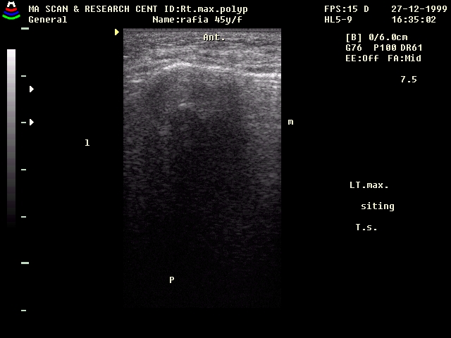
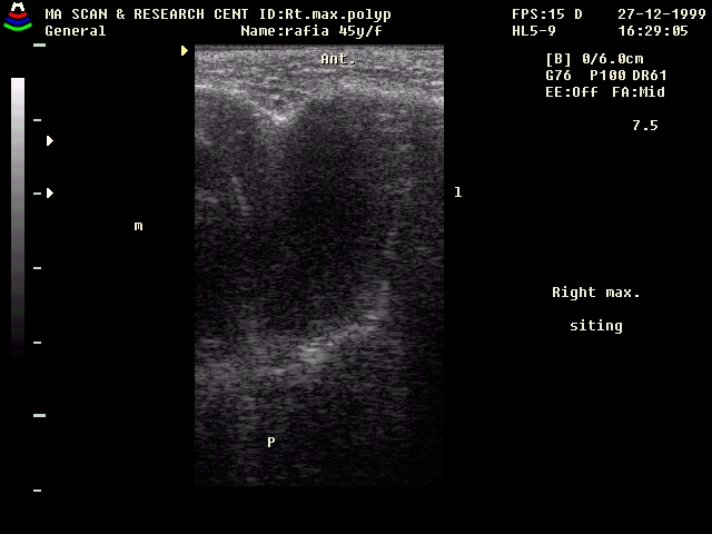
EXAMPLE TWO : Lt.maxillary polyposis.
|
|
||||||||||
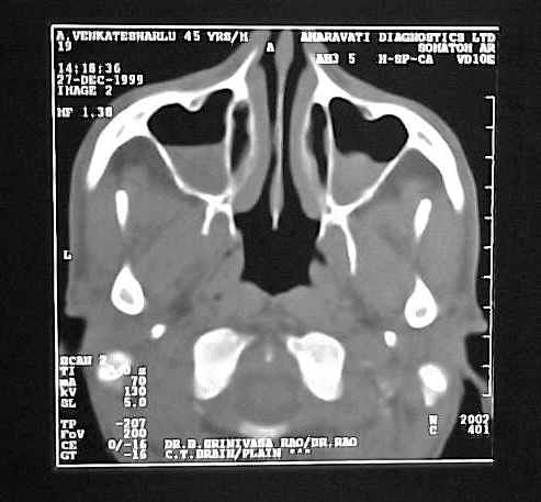
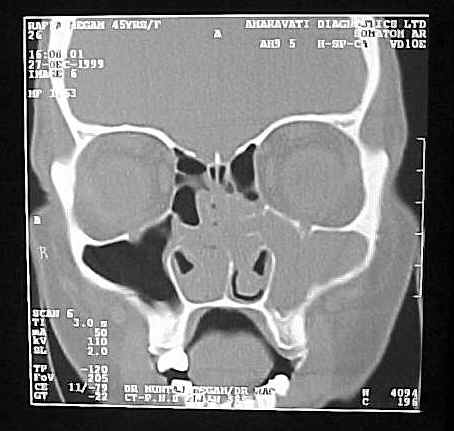
| Ultrasonography of lt.maxillary shows hypoechoic cavity with echogenic bony walls.(apex,lateral & medial walls) | LT.Maxillary sinus is filled with hypodense area with intact bony walls. |
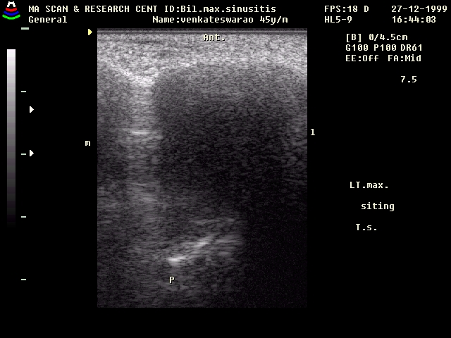
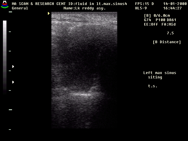
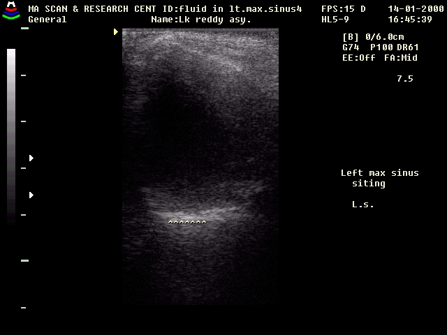
EXAMPLE THREE : FLUID IN MAXILLARY SINUS.
|
|
|
| Lt.maxillary sinus (T.S & L.S. in siting position)Apex of maxillary sinus is seen very well. But medial & lateral
walls are not seen.
|
Air-fluid level. |
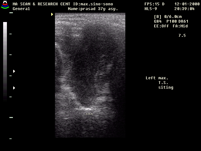
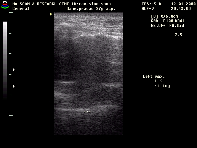
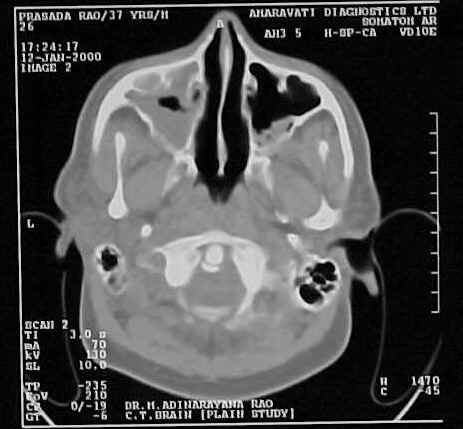
EXAMPLE FOUR : LT.MAXILLARY MUCOSAL THICKENING
37 Y Male pateint complaining of headache at vertex. No h/o bouts
of sneezings.
|
|
|
| LT.maxillary sinus (T.S.in siting position)):Mucosal thickening well seen along the lateral wall.
Apex ,medial, lateral wall are well seen.Sinus cavity is hypoechoic.
|
LT.maxillary sinus (L.S.in siting position) : Mucosal thickening is well seen in the inferior & anterior wall. |
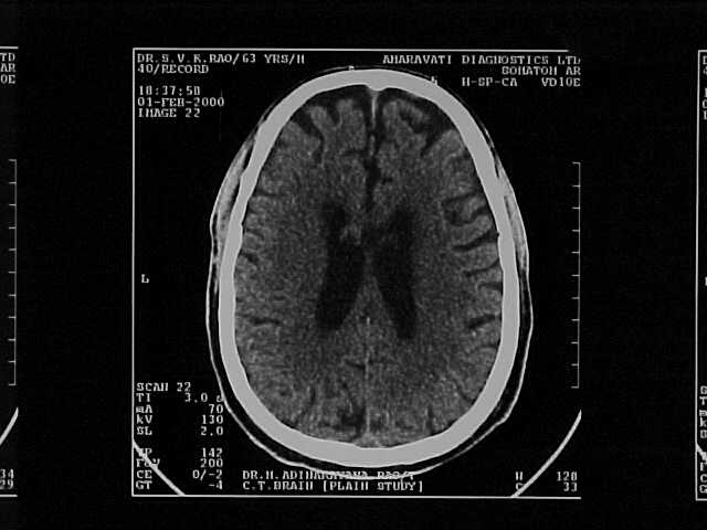
CT SCAN shows mucosal thickening in lt.maxillary sinus. |
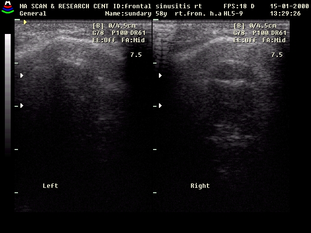
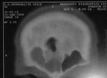
RT.FRONTAL SINUSITIS:
|
|
| Lt. frontal sinus shows hyperechoic bony wall. Contents of
cavity are not seen .Posterior bony wall is not seen.
Rt.frontal sinus shows hyperechoic bony wall . Contents of cavity are hypoechoic.Posterior bony wall is seen. |
CT scan shows rt.frontal sinusitis. Lt.maxillary sinus is normal. |
CONCLUSIONS :
High resolution
sonography is highly sensitive & specific in expert hand to detect maxillary
sinus pathology like mucosal thickning and fluid fluid layer. And is cost
effective easly available compare to CT scan. Lack of radiation & real time
scanning are boon with ultrasonography.
Ultrasound is also useful to detect frontal sinusitis.